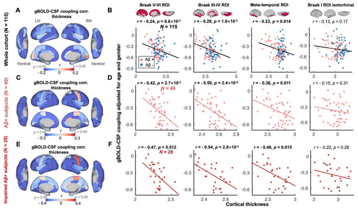
Fig. 2. gBOLD-CSF coupling is correlated with cortical thickness across the whole cohort and Aβ+ subjects.
(A) Subjects with weaker (less negative) gBOLD-CSF coupling had thinner cortices in the majority of brain regions, especially in the frontal, parietal, and temporal lobes, including default mode network (DMN) and fronto-parietal network (FPN). (B) The gBOLD-CSF coupling strength significantly decreased with thinner cortices in Braak V-VI, Braak III-IV, and temporal meta-ROIs across the whole cohort (all r < − 0.23, all p < 0.014; N = 115). A similar but not significant coupling-thickness association was found in the entorhinal region (r = − 0.13, p = 0.17). (C-F) Among Aβ+ subjects, particularly impaired Aβ+ ones, the coupling-thickness remained striking (all r < − 0.36, all p < 0.015 in Braak V-VI, Braak III-IV, and temporal meta-ROI in D and F) while this was not the case in other participants (Fig. S4). Each point represents one subject.

