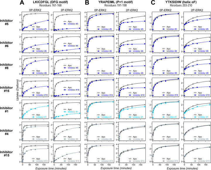
Figure 4. HDX assays survey conformation selection among compounds in the ERK inhibitor panel.
HDX measurements performed with representative inhibitors shown in Fig. 3, chosen for variations in their left-side, central scaffold, and right-side substituents. Time courses show deuterium uptake at the (A) DFG motif (peptide 161–168: LKICDFGL), (B) P+1 segment (peptide 191–198, YRAPEIML), and helix αF (peptide 203–210: YTKSIDIW). Enhanced HDX protection (strongly decreased uptake) in each segment by inhibitors #5, #6, #8 and #16 (blue) suggest properties of conformation selection for the R-state, while lower protection by inhibitors #4 and #15 (grey) suggest retention of conformational exchange. Inhibitor #1 (cyan) shows HDX properties intermediate to these two groups. HDX time courses for the full set of 17 inhibitors are shown in Suppl. Fig. S6A,B.

