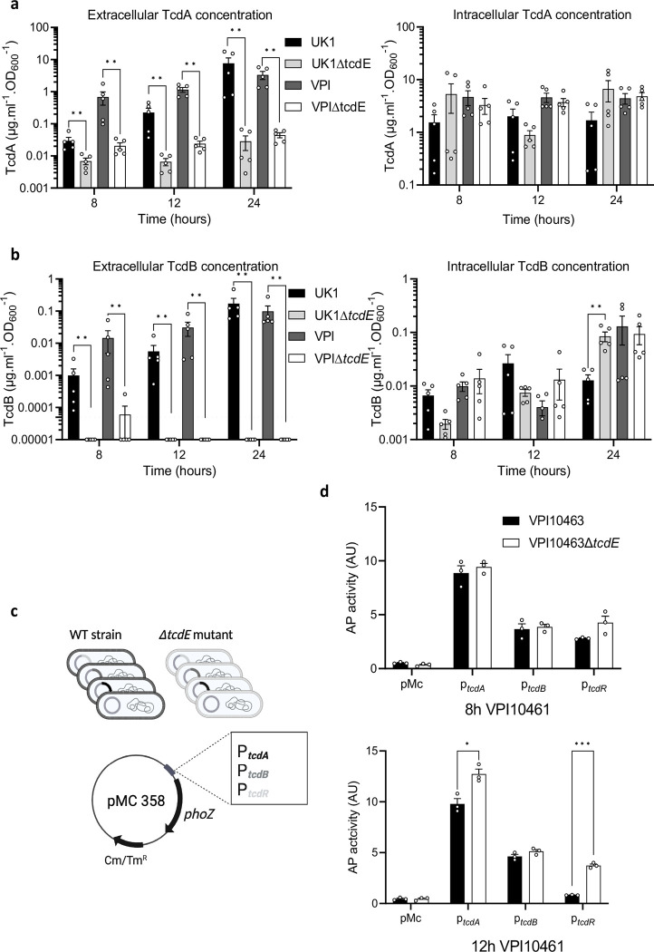
Fig. 2. TcdE mediates TcdA and TcdB release in VPI10463 and UK1 strains in vitro.
a. and b. TcdA (a) and TcdB (b) titers in extracellular (left panel) and intracellular (right panel) fractions of VPI10463 and UK1strains and their respective ΔtcdE mutants, after 8, 12 and 24 hours of growth. Strains were grown in TY medium, and toxins were quantified using TcdA- and TcdB-ELISA. Means and SEM are shown; n=5 independent experiments. ** p<0,01 by a Mann-Whitney test. Horizontal dotted line shows thresholds of detection. c. Schematic representation of transcriptional fusions constructions. Transcriptional fusions of promoter regions of approximately 500 bp of tcdA, tcdB or tcdR genes fused to the reporter gene phoZ, were introduced by conjugation into the VPI10463 wild type strain and the isogenic ΔtcdE mutants. d. Alkaline phosphatase (AP) activity of PtcdA::phoZ, PtcdB::phoZ and PtcdR::phoZ fusions expressed from pMC358 in VPI10463 and VPI10463ΔtcdE. Strains were grown in TY medium and samples assayed for AP activity were collected at 8 and 12 hours of growth. Means and SEM are shown; n=3 independent experiments. *P ≤ 0.05 and ***P ≤ 0.001 by an unpaired t test.

