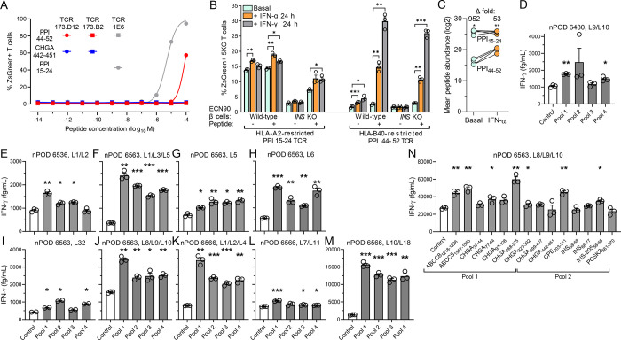
Figure 6. Recognition of HLA-B40-restricted peptides in the islets of T1D donors.
A. Dose-response peptide recall of 173.D12, 1E6 and negative control 173.B2 TCR-transduced ZsGreen-NFAT reporter 5KC T-cells co-cultured for 18 h K562 antigen-presenting cells transduced with HLA-B40 (for 173.D12 and 173.B2) or HLA-A2 (for 1E6) and pulsed with the indicated peptides. A representative experiment out of 2 performed is shown. B. Activation of ZsGreen-NFAT reporter 5KC T-cells transduced with a 1E6 TCR recognizing HLA-A2-restricted PPI15–24 or a 173.D12 TCR recognizing HLA-B40-restricted PPI44–52. Following the indicated cytokine pretreatment, ECN90 β-cells (wild-type or INS KO) left unpulsed or pulsed with the cognate peptide were put in contact with TCR-transduced 5KC T-cells for 6 h. Data represent mean±SEM of triplicate measurements from a representative experiment performed in triplicate. *p<0.05, **p<0.01 and ***p<0.001 by Student’s t test. C. Average total abundance and fold difference of HLA-A2-restricted PPI15–24 and HLA-B40-restricted PPI44–52 peptides presented under basal and IFN-α-treated conditions (n=4/each; PPI15–24 but not PPI44–52 was detected in 4 additional replicates). *p<0.05 and **p<0.01 by paired Student’s t test. D-N. IFN-γ secretion by polyclonal CD8+ T-cell lines expanded from islet infiltrates of HLA-B40+ nPOD T1D donors (listed in Supplementary Table 2) and exposed to HLA-B40-transduced K562 antigen-presenting cells pulsed with HLA-B40-restricted peptide pools (D-M; listed in Supplementary Table 3) or with individual peptides (N). Data represent mean±SEM of triplicate measurements from a representative experiment performed in duplicate. *p<0.05, **p<0.01 and ***p<0.001 by paired Student’s t test.

