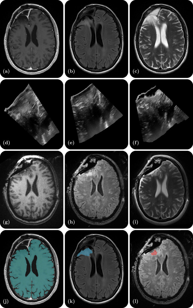
Figure 1.
Illustrative example of one dataset - a right frontal lobe recurrent WHO Grade II Oligodendroglioma (IDH-positive, 1p/19q co-deleted). (a) Preoperative contrast-enhanced T1-weighted MR; (b) Preoperative T2-weighted MR; (c) Preoperative T2 FLAIR MR; (d) Intraoperative US prior to dural opening; (e) Intraoperative US post dural opening; (f) Intraoperative US prior to iMRI; (g) Intraoperative contrast-enhanced T1-weighted MRI; (h) Intraoperative T2 FLAIR MRI; (i) Intraoperative T2-weighted MRI (BLADE); (j) Cerebrum segmentation on the preoperative contrast-enhanced T1-weighted MRI; (k) Tumor segmentation on the preoperative T2 FLAIR MRI; (l) Residual tumor segmentation on the intraoperative T2 FLAIR MRI.

