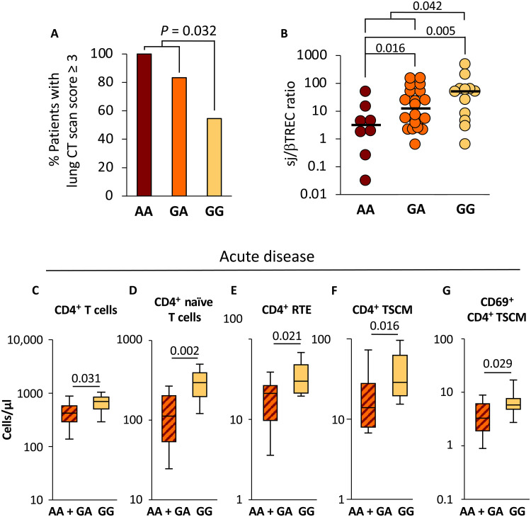
Fig. 1. Patients with GG genotype displayed less severe pulmonary involvement and higher thymic output.
Patients with COVID-19 were classified according to their genotype at the rs2204985 (n = 8 AA, n = 20 GA, and n = 12 GG) locus and plotted as a function of pulmonary involvement severity (A) and thymic function (sj/βTREC ratio; B). Total CD4+ T cells, naïve (CD45RA+, CD95−, CD27+, CD28+) CD4+ T cells, CD4+ RTE (CD45RA+, CD95−, CD27+, CD28+, CD31Hi), CD4+ TSCM (CD45RA+, CD95+, CD27+, CD28+), and activated (CD69+) CD4+ TSCM were quantified by fluorescence-activated cell sorting in peripheral blood mononuclear cells (PBMCs) from patients with COVID-19 sampled during the acute phase of the disease (C to G). Statistical significance of the differences between groups is shown on top [logistic regression model for (A) and Mann-Whitney test for (B) to (G)]. Patients with AA and GA genotypes were analyzed in the same group. The gating strategy for T cell subsets is shown in fig. S1.

