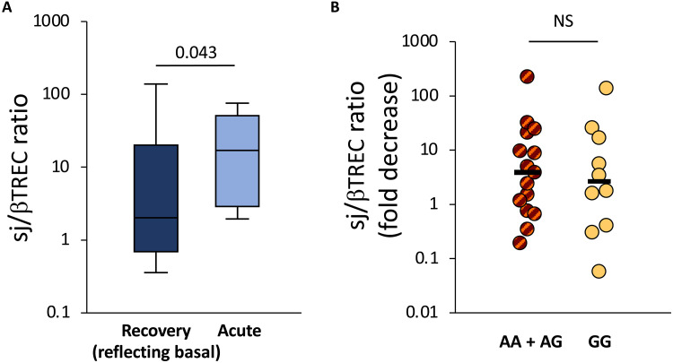
Fig. 2. Thymic function during acute disease and after recovery from COVID-19.
Thymic function was assessed through the quantification of the sj/βTREC ratio in patients with COVID-19 during their first hospitalization in ICU and 6 months after recovery are shown (A). The fold decrease in sj/βTREC ratio between samplings for AA + GA and GG patients is shown (B). Horizontal bars represent medians in each patient group. Statistical differences between AA + GA and GG patients at each time points (Mann-Whitney test for two independent samples). The recovery phase is expected to reflect some features present during the basal conditions. NS, not significant.

