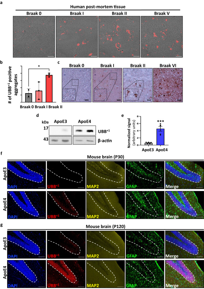
Fig. 1. UBB+1 accumulates in the early stages of AD postmortem tissue.
a Immunofluorescence staining of human tissue (ER = hippocampal complex, including entorhinal cortex) with an antibody against UBB+1 (red). b Quantification of UBB+1-positive aggregates counted in different biological samples taken from dentate gyrus (DG) of patients diagnosed at different Braak stages [n = 3 biologically independent samples] (Supplementary information Table 1) (p = 0.038). c Immunohistochemistry staining of human postmortem tissue (ER) with anti-UBB+1. d Representative immunoblots of proteins isolated from the hippocampus of Apolipoprotein E (ApoE3/4)-TR mice using an anti-UBB+1 antibody. e Quantification of (d) by densitometry and normalized to β-actin [n = 5 mice] (p = 0.00042) f, g Immunofluorescence staining of hippocampal sections of one (f) and 4-month-old (g) ApoE (ApoE3/4)-TR mice, showing UBB+1 is specifically expressed in neurons (MAP2) and not glial cells (GFAP). P-values were determined by unpaired two-tailed Student’s t-test. Error bars represent ± s.d. Images are representative of three independent wells. All experiments were repeated at least twice.Scale bars: 20 µm (a), 50 μm (c), 100 μm (f, g).

