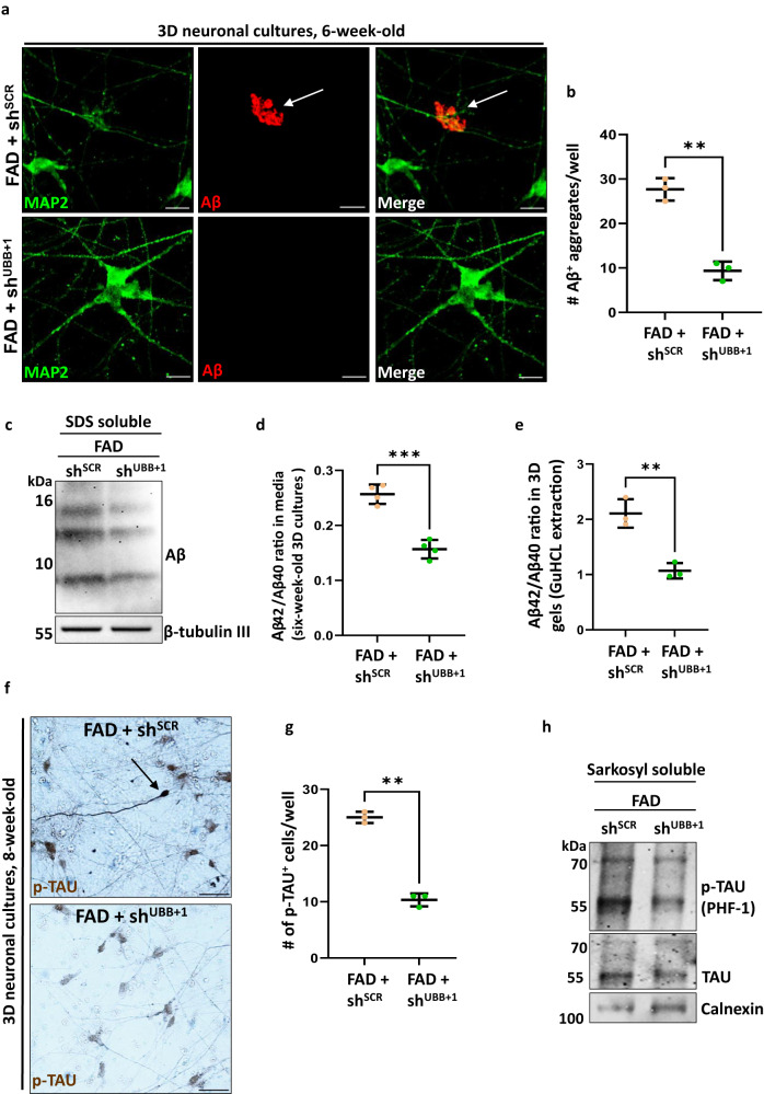
Fig. 4. Silencing UBB+1 expression decreases Aβ and p-tau aggregates in a 3D human neuronal culture.
a Immunofluorescence of Aβ deposits in 6-week- old FAD+shSCR or FAD+shshUBB+1 (green, MAP2; red, 3D6; arrowheads, extracellular Aβ deposits). b Quantification of extracellular Aβ deposits in FAD+shSCR or FAD+shshUBB+1 cultures [n = 3 biologically independent samples, the whole well was counted] (p = 0.0006)c Immunoblot of Aβ aggregates from 6-week-old FAD+shSCR or FAD+shshUBB+1 cultures. d Quantification of Aβ42/Aβ40 by ELISA of conditioned media from 6-week-old FAD+shSCR or FAD+shshUBB+1 cultures [n = 4 biologically independent samples] (p = 0.00018). e Quantification of Aβ42/Aβ40 by ELISA of lysates from 6-week-old FAD+shSCR or FAD+shshUBB+1 cultures [n = 4 biologically independent samples] (p = 0.0373). f Immunohistochemistry of 8-week-old FAD+shSCR or FAD+shshUBB+1 cultures showing p-tau staining (brown, p-tau; arrows indicate cells with high levels of p-tau). g Quantification of p-tau deposits in 8-week-old FADSCR or FADshUBB+1 cultures. [n = 3 biologically independent samples, the whole well was counted] (p = 0.00007). h Immunoblot of 8-week-old FAD+shSCR or FAD+shshUBB+1 cultures using anti-p-tau, and anti-tau, Calnexin was used as housekeeping gene. P-values were determined by unpaired two-tailed Student’s t-test where. Error bars represent ± s.d. Images are representative of three independent wells. All experiments were repeated at least twice. Scale bars: 20 µm (a), 50 µm (f).

