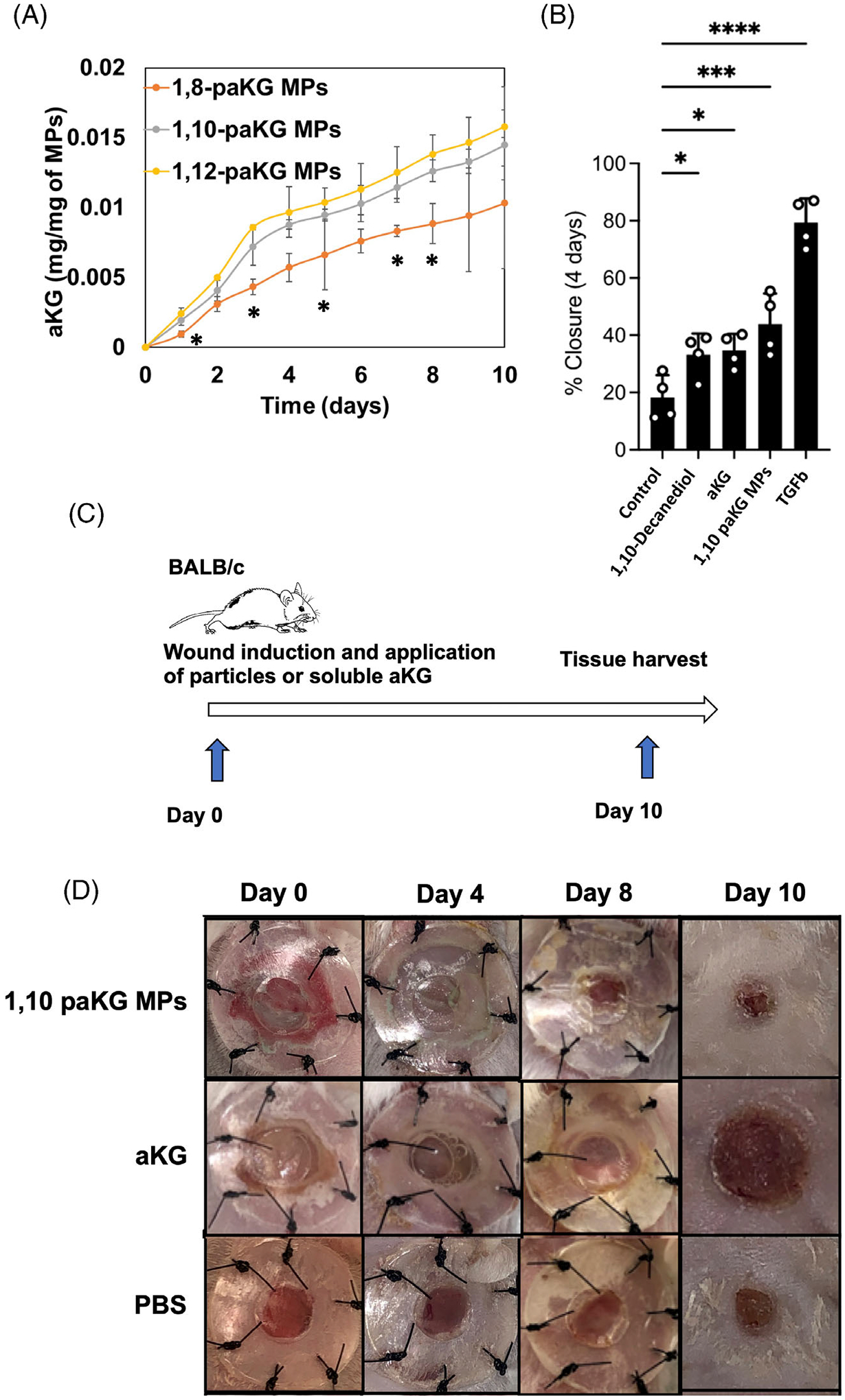
FIGURE 2.

paKG MPs facilitate faster wound closures in vitro and in vivo. (A) Release kinetics of aKG was performed from the microparticles generated from different polymers (n = 3, avg ± SD, *p < .05 significance of 1,8-paKG MPs as compared to 1,10-paKG MPs and 1,12-paKG MPs, Student’s T test). (B) Percentage closure of the wound (scratch assay in HaCaT human keratinocyte cells in vitro) was quantified on day 4, which demonstrates that 1,10-paKG MPs were able to decrease the wound area significantly higher than the PBS control (n = 4, avg ± SEM, *p < .05, One-way ANOVA). (C) In vivo study design. (D) Representative images of the wound after treatment with 1,10-paKG microparticles demonstrate faster wound healing as compared to the other groups. (Note – the dark region represents scab and closed wound in 1,10-paKG microparticles group) (n = 6, avg ± SEM, *p < .05).
