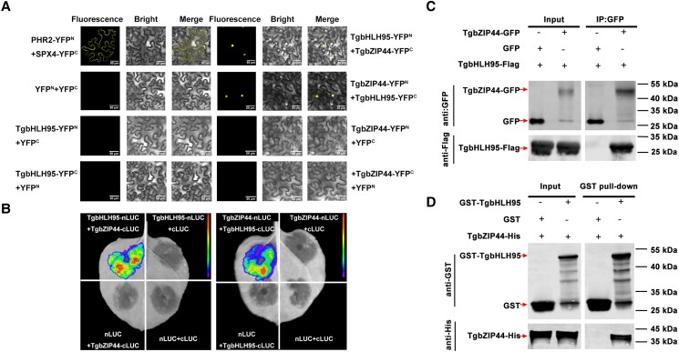
Figure 6.
TgbHLH95 protein interacts with TgbZIP44. A) BiFC analysis of the interaction between TgbHLH95 and TgbZIP44. The pairs of fusion proteins tested were TgbHLH95-YFPN + TgbZIP44-YFPC and TgbZIP44-YFPN + TgbHLH95-YFPC. PHR2-YN and SPX4-YC were used as the positive control; YFPN and YFPC were used as negative controls. Nuclear location signal (NLS) indicates the position of the nucleus. Scale bars = 50 µm. B) LCI analysis of the interaction between TgbHLH95 and TgbZIP44. Tobacco leaves were infiltrated with Agrobacterium strain GV3101 harboring different constructs. The luminescence signals of the infiltrated areas were measured 48 h after infiltration. The experiment was repeated at least 3 times. C) Co-IP assay of TgbHLH95 and TgbZIP44. TgbHLH95-Flag plus TgbZIP44-GFP proteins were immunoprecipitated with an anti-GFP antibody and immunoblotted with anti-GFP and anti-Flag antibodies. D) Pull-down assay detection of the interaction of TgbHLH95 and TgbZIP44. The recombinant TgbHLH95-GST or GST was incubated with TgbZIP44-His. Blots were probed with anti-GST and anti-His antibody.

