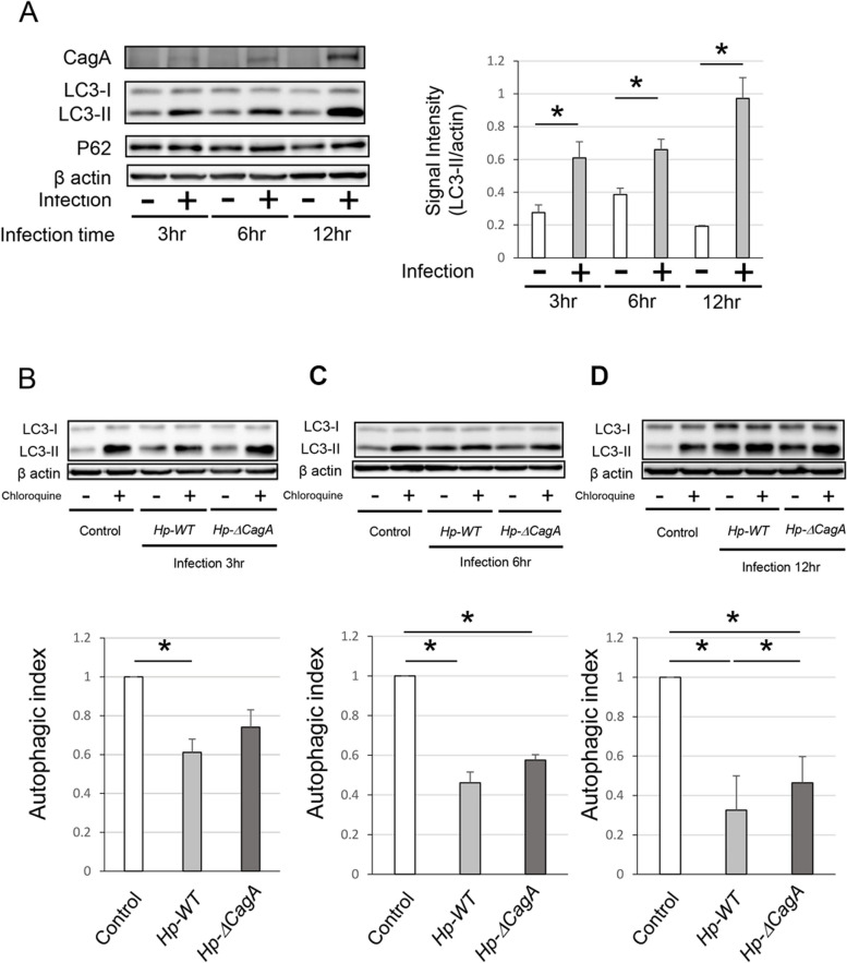
Fig. 1.
Autophagy in AGS cells was inhibited after infection with Helicobacter pylori. A Western blotting showing the protein levels of CagA, LC3, and P62 in AGS cells infected with Hp-WT for 3, 6, and 12 h (n = 5, mean ± SD, *P < 0.05). B-D) Western blotting and autophagic flux assays in AGS cells infected with Hp-WT and Hp-ΔcagA for 3(B), 6(C), and 12 h(D) (n = 5, mean ± SD, *P < 0.05). CagA, cytotoxin-associated gene A; LC3, microtubule-associated proteins 1A/1B light chain 3A; Hp-WT, wild-type cagA-positive H. pylori; Hp-ΔcagA, cagA-knockout H. Pylori; SD, standard deviation

