Figure 2.
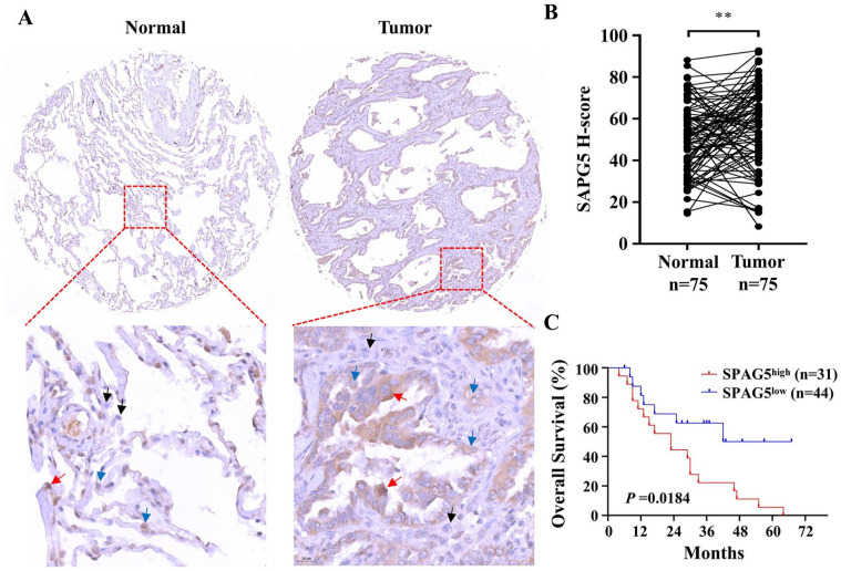
TMA IHC staining reveals elevated expression of SPAG5 in LUAD tumor tissue, and its upregulation correlates with unfavorable overall survival. (A) Representative images of SPAG5 IHC staining in LUAD TMA are presented, with the lower panel displaying a magnified view of the region highlighted by the red dashed box in the upper panel. Black, blue, and red arrows indicate cells with negligible expression, moderate expression, and high expression of SPAG5, respectively. (B) H-score analysis of IHC staining demonstrates significantly higher SPAG5 expression in LUAD tumor tissue (n = 75) compared with normal tissue (n = 75). (C) The Kaplan-Meier survival curve, analyzed using the log-rank test, illustrates a significant association between elevated expression of SPAG5 and a notably poorer prognosis (P = .0184). IHC indicates immunohistochemistry; LUAD, lung adenocarcinoma; SPAG5, sperm-associated antigen 5; TMA, tissue microarray.
**P < .01.

