Fig. 7.
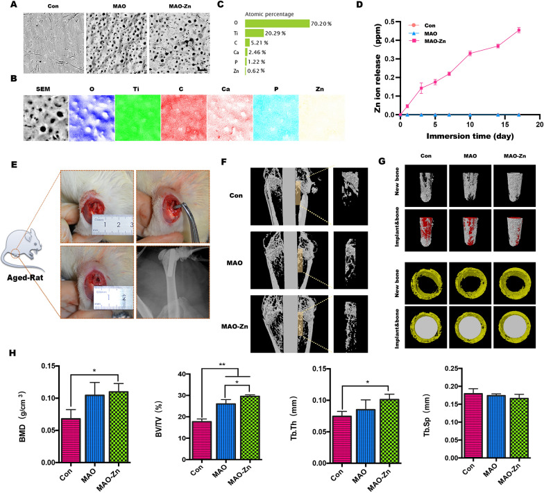
Analysis of the products after knee implantation in aged rats. a Scanning electron microscopy images of the implant surfaces. b, c Energy-dispersive X-ray spectroscopy analysis of the micro-arc oxidation implant with zinc acetate addition to the electrolyte (MAO-Zn2+) group. d Cumulative concentrations of Zn.2+ released from MAO-Zn during the immersion in a PBS solution at 37 °C. e Model diagram and X-ray image of the metal implant model of the aging rat femur. f, g Promoting osteointegration in vivo detected by microCT in 2D or 3D views. h Statistical analysis of bone mineral density (BMD), bone volume to tissue volume (BV/TV), trabecular thickness (Tb.Th), and trabecular separation (Tb.sp) using the acquired microCT data for each group (n = 3, *p < 0.05, **p < 0.01)

