
Fig. 1.
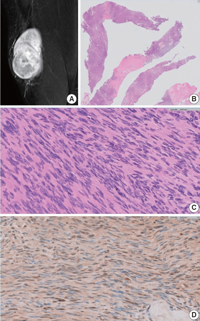
A cellular schwannoma. (A) Magnetic resonance imaging scan showing a heterogeneously enhancing mass within the subcutaneous tissue plane. (B) Core biopsies showing predominantly cellular areas, resulting in a “blue appearance” accompanied by pale degenerate areas. (C) Compact spindle cells showing hypercellularity and mild nuclear pleomorphism. (D) S-100 immunohistochemistry showing a focal weak staining pattern despite high cellularity.