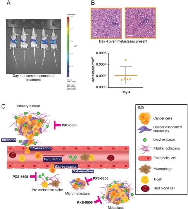
Extended Data Fig. 7. Liver colonization in the intrasplenic model and schematic of PXS-5505 inhibition of lysyl oxidases in PDAC.
a. IVIS imaging of late-stage treatment study of liver colonization model over 11 days. b. Representative images of H&E-stained liver at day 4 confirming presence of micro-metastatic lesions as detected by IVIS imaging n = 5 independent animals shown in (A). Data presented as mean values +/− SD. Scale bar 100 μm. Data also shown in Fig. 5f (orange) for reference b. PXS-5505 inhibits lysyl oxidase family member activity at different stages of tumor progression and metastasis.

