Fig. 2.
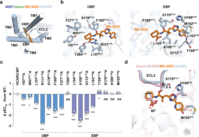
Key residues for the interaction between MK-6892 and HCAR2. a Structural comparison of the binding modes of niacin, MMF and MK-6892 to HCAR2. b The detailed interactions between MK-6892 and HCAR2 in OBP (left panel) and EBP (right panel). Black dashed lines represent polar interactions. c Mutagenesis effects of the residues in OBP and EBP of HCAR2 on their activities in response to niacin and MK-6892 stimulation examined by cAMP inhibition assay. The value of ΔpEC50 (pEC50MT-pEC50WT) shows differences between wild-type (WT) receptors and mutants (MT). Data are displayed as mean ± SEM from at least three independent experiments, each performed in triplicate. Statistical significance was determined by one-way analysis of variance with Dunnett’s multiple comparison test (compared to WT). n.s., no significance. d Structural comparison of the niacin- and MK-6892 bound binding pocket in HCAR2. The conformational changes between the two structures are indicated as red arrows

