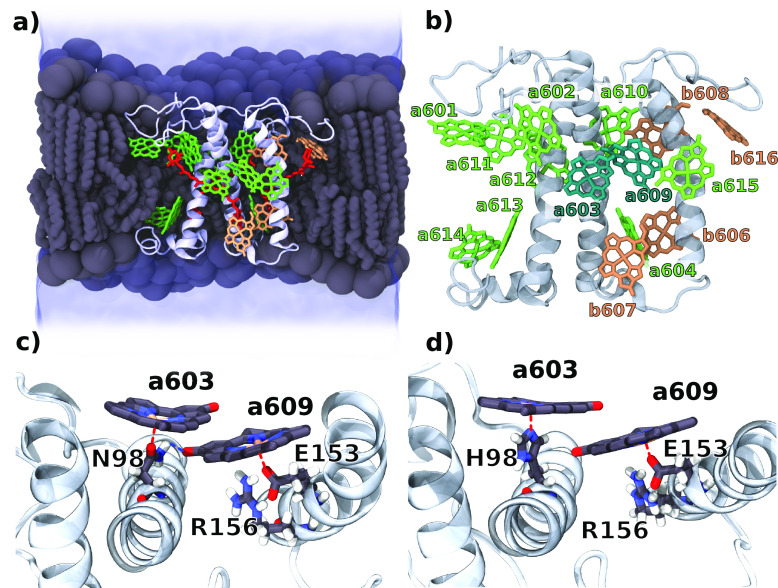
Figure 1.
Molecular structure of Lhca4. (a) Longitudinal section of the simulated system. The Lhca4 complex was embedded in the phospholipid bilayer (gray) and water solution (blue). Pigment composition and protein orientation in the membrane are highlighted in the picture. Chls a are represented in green, Chls b in orange, and carotenoids in red. (b) Chl positions on the Lhca4 complex. The dimer a603–a609 is highlighted. (c) Binding pocket of Chls a603–a609 in the WT with highlighted binding residues. (d) Binding pocket of Chls a603–a609 in N98H. The red dashed lines represent coordination to the Mg atom, while the blue dashed line represents the hydrogen bond between Asn and Chl a609.

