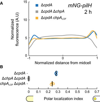
Figure 3. ChpA promotes PilH localization in a phosphorylation‐independent manner.

- mNG‐PilH fluorescence profiles in chpA mutants after 2 h surface growth. To avoid negative effects of low cAMP level on localization of PilH, cpdA was deleted in all displayed strains to rescue cAMP level to WT levels (cf. Appendix Fig S4B). Solid lines, mean normalized fluorescence profiles across biological replicates. Shaded area, standard deviation across biological replicates.
- PilH polar localization is abolished in ΔchpA. PilH polar localization is however maintained in chpA LOF . Circles, median of each biological replicate. Vertical bars, mean across biological replicates. For corresponding asymmetry indexes and mean cell fluorescence, see Appendix Fig S2C.
