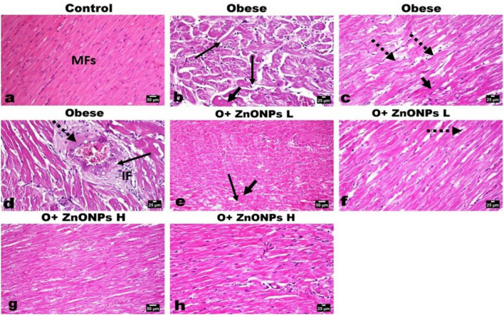
Figure 9.
Photomicrographs of H&E-stained heart sections of various experimental groups. (a) Heart of control rat showing normal orientation ad striation of cardia muscles’ fibers (MFs). (b–d) Heart of obese rat showing marked degeneration (arrow), loss of striation, and scattered eosinophilia (short arrow) of the myofibers with increase intermuscular fat (dotted arrow). (d) The coronary vessels showing vacuolization of medial muscles (arrow) and focal fibroid necrosis (dotted arrow), and inflammatory cells infiltration (IF). (e, f) Heart of low dose ZnONPs administrated rat showing moderate degree of myofibers degeneration (arrow), mild eosinophilia (short arrow) with some intermuscular fat (dotted arrow). (g, h) Heart of high dose ZnONPs administrated rat showing good restoration of the cardiac muscle fibers with mild degeneration (arrow) and decreased intermuscular fat.

