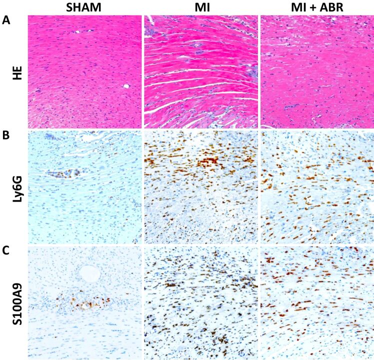
Figure 1.
Neutrophil infiltration and S100A9 presence in the myocardium on day 1 post-MI: (A) Representative images of HE staining of left ventricular sections from the sham and MI groups treated with PBS (MI) or ABR-238901 (MI+ABR), respectively, collected on day 1 after myocardial ischemia (400×); (B) Ly6G-positive staining identifying neutrophil infiltration (400×); (C) S100A9-positive immunohistochemical staining in the same areas as in (A) and (B) (400×). HE: Hematoxylin–Eosin; Ly6G: Lymphocyte antigen 6 family member G6D; MI: Myocardial infarction; PBS: Phosphate-buffered saline

