Figure 1.
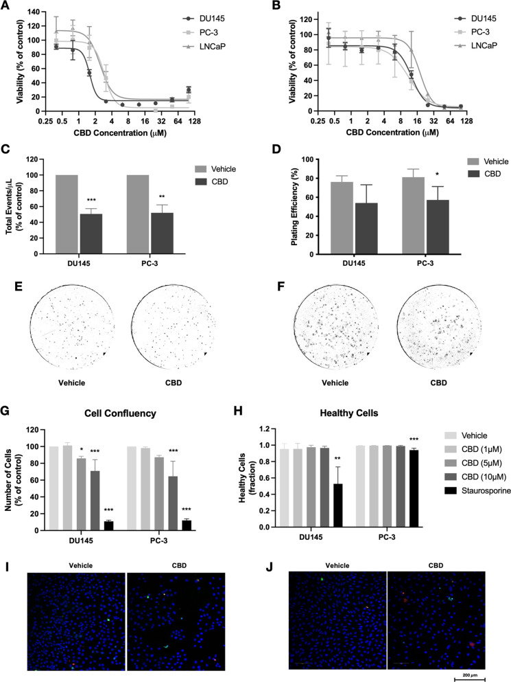
CBD reduces the viability, survival, and proliferation of prostate cancer cells. (A) Prostate cancer cells (DU145, PC-3, LNCaP) were treated with CBD (0–100 μM) for 72 h in the absence of serum. Cell viability was measured using the MTT assay. (B) Prostate cancer cells were treated with CBD in the presence of 10% FBS. Cell viability was measured using the MTT assay. (C) Androgen-insensitive cells (DU145, PC-3) were treated with IC50 doses of CBD for 48 h. Cell counts were determined using flow cytometry. (D) Cells were treated with IC50 doses of CBD for 48 h before reseeding without treatment. Colonies formed were counted after 7 days. (E) Representative images of colony formation in DU145 cells. (F) Representative images of colony formation in PC-3 cells. (G) Cells were treated with CBD (1, 5, 10 μM) for 72 h. Cell confluency was assessed by high-content fluorescence microscopy using Hoechst 33342 staining. Staurosporine was used as a positive control. (H) The fraction of healthy cells was measured using YO-PRO and propidium iodide (PI) staining. (I) Representative images of DU145 cells treated with vehicle or 10 μM CBD, stained with Hoechst 33342 (blue), YO-PRO (green), and PI (red). (J) Representative images of PC-3 cells treated with vehicle or 10 μM CBD. Data are represented as mean ± SD calculated from at least three independent experiments. *p < 0.05, **p < 0.01, ***p < 0.001 compared to the vehicle control for that cell line.

