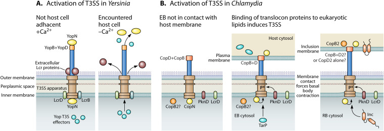
FIG 3.
Activation of T3SS. (A) Depicts how the Yersinia T3SS is activated by host cell contact, which was originally characterized by chelating Ca2+ from bacterial growth medium (127, 256–258). (B) Depicts a likely mechanism of chlamydial T3SS activation, which is contact with lipids of the plasma membrane or the inclusion membrane (145). This model further depicts possible differences in the translocon proteins of an EB (CopB and CopD) versus an RB (CopD2 only or CopB and CopD2). CopB2 is modeled as a peripheral inclusion membrane protein as is consistent with current data (123).

