Fig. 1.
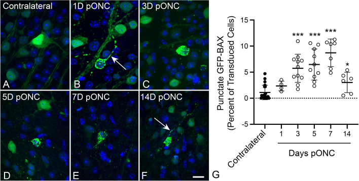
Static imaging of GFP-BAX expressing cells in BALB/cByJ mice showing the time course for GFP-BAX translocation as a function of time after optic nerve crush (ONC). Previously, we had documented that the translocated GFP-BAX forms puncta localized to mitochondria in these cells [25]. A-F Static imaging of GFP-BAX expressing cells in retinal whole mounts at times post-ONC (pONC) showing localization changes from diffuse to punctate distribution of the fusion protein. In some cells, punctate GFP-BAX is also evident in the axon originating from the cell (arrows in B, F). Scale bar = 20 µm. G Graph showing the change in percentage of GFP-BAX transduced cells that exhibit punctate labeling pONC. A significant increase in punctate cells is evident at 3, 5, 7 and 14 days pONC relative to contralateral eyes (*P = 0.012, *** P < 0.0001, individual t-tests). This pattern is consistent with descriptions of BAX accumulation in cells of the ganglion cell layer after optic nerve damage in rats and mice reported by others [25, 32, 48, 50]

