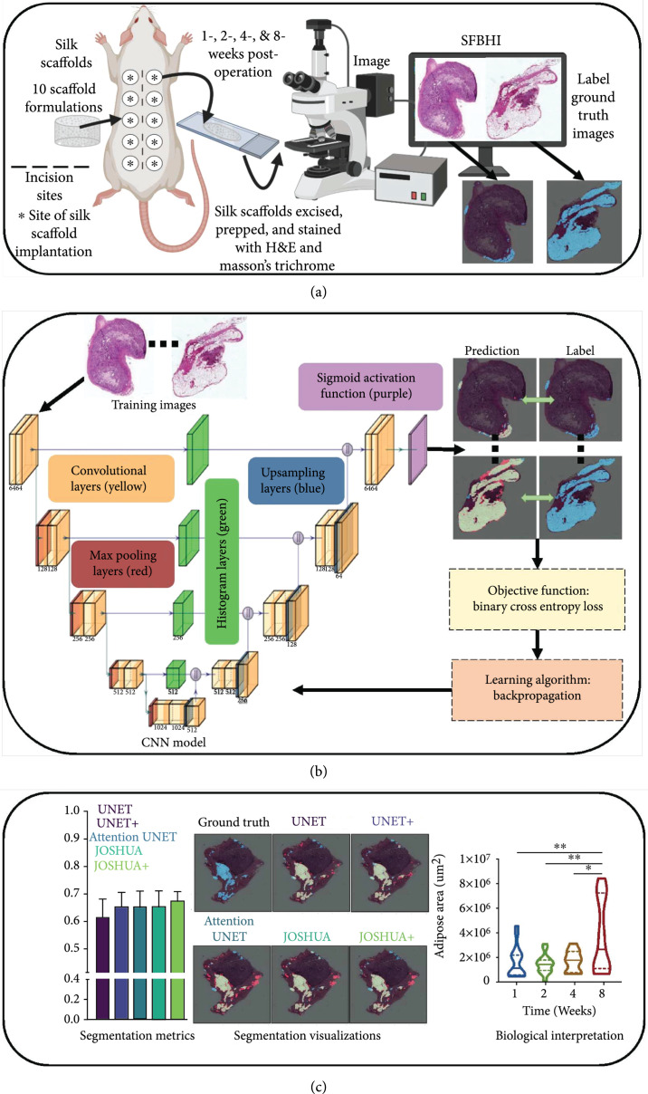
Figure 1.
Our overall process has three components: data collection, processing, and analysis as shown in Figures 1(a) through 1(c), respectively. (a) For data collection, acellular silk fibroin-lyophilized sponges of varying formulations were subcutaneously inserted into the lateral pockets of Sprague Dawley rats. After 1-, 2-, 4-, and 8-week postsurgery, the silk sponges and overlaying tissue were excised. The samples were prepped and stained with H&E and Masson’s trichrome. (b) The next step (i.e., data processing) trains our proposed model through -fold cross validation. (c) Once training is completed, we can use our machine learning models to quickly segment and quantify the adipose tissue for new samples. We then perform data analysis to connect the model outputs to meaningful biological interpretations.

