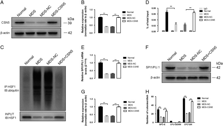
FIGURE 2.
Effect of CSN5 overexpression in CD34+ cells of MDS patients. A, The protein expression of CSN5 in whole cell lysates of CD34+ cells of normal, MDS, MDS-NC, and MDS-CSN5. B, β-actin was used as the loading control for the whole cell lysate. Densitometry represents the expression of the proteins relative to β-actin. C, The results obtained by coimmunoprecipitation, and HSF1 ubiquitination levels were measured with western blot assay. Densitometry represents the expression of the proteins relative to input. D, ChIP assay shows HSF1 binding to PU.1/Spi1 promoter. The ChIP ratio of PU.1/Spi1 relative to input. E, PU.1/Spi1 mRNA levels were detected by real-time PCR. And GAPDH was used as the loading control. F, The protein expression of PU.1/Spi1, in whole cell lysates of CD34+ cells of normal, MDS, MDS-NC, and MDS-CSN5. G, β-actin was used as the loading control for the whole cell lysate. Densitometry represents the expression of the proteins relative to β-actin. H, The formation of BFU-E, CFU-GEMM, and CFU-GM was used with colony formation assay. All data are shown as mean ± SD (n = 3). *P < 0.05; **P < 0.01.

