. 2016 Nov 7;60(6):587–595. doi: 10.1590/2359-3997000000230

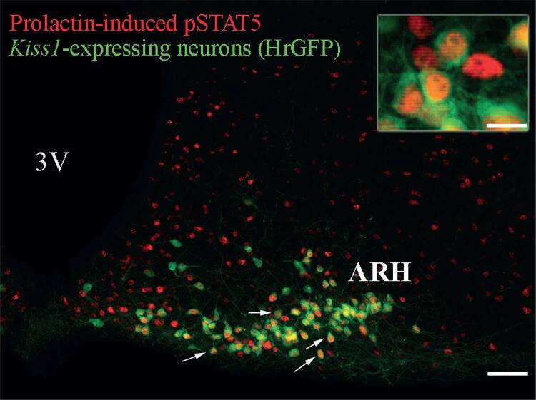
Figure 2. Prolactin-responsive Kiss1-expressing neurons in the mouse arcuate nucleus. Fluorescent photomicrographs of hypothalamic sections showing the expression of humanized Renilla green fluorescent protein (hrGFP) which is expressed under the transcriptional control of the Kiss1 gene and prolactin-induced pSTAT5 immunoreactivity of a female mouse in diestrus. Details about this mouse model can be found in previous publications (Cravo and cols., 2013). Mice received a single i.p. injection of ovine prolactin (10 µg/g) before perfusion. Arrows illustrate examples of prolactin-responsive Kiss1-expressing neurons. The inset represents a higher magnification of dual-labeled neurons in the ARH. Abbreviations: 3V, third ventricle. Scale bar: photomicrograph = 100 µm; inset = 23 µm.

Figure 2