. 2019 Nov 1;63(6):549–556. doi: 10.20945/2359-3997000000184

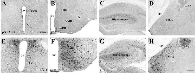
Figure 1. Distribution of GH responsive cells in different brain areas of mice via the detection of STAT5 phosphorylation (pSTAT5). A-H. Brightfield photomicrographs of brain sections showing the distribution of pSTAT5 immunoreactive cells in saline-injected mice (A-D) or in GH-injected mice (E-H). Mice received an intraperitoneal injection of 20 µg of porcine GH per gram of body weight, followed by perfusion 90 minutes later. Abbreviations: 3V, third ventricle; ARH, arcuate nucleus; CEA, central nucleus of the amygdala; DMH, dorsomedial nucleus; fx, fornix; MEA, medial nucleus of the amygdala; opt, optic tract; PVH, paraventricular nucleus; VMH, ventromedial nucleus. Scale Bar = 200 µm. Figure adapted from the study by Furigo and cols. (47).

Figure 1