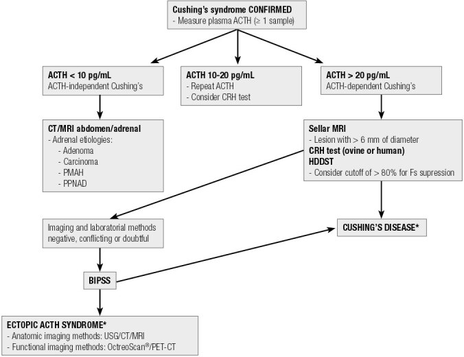
Figure 2. Flowchart for differential diagnosis of ACTH-dependent Cushing’s syndrome.

HDDST: high-dose dexamethasone suppression test (8 mg overnight); CT: computed tomography; MRI: magnetic resonance imaging; PMAH: primary macronodular adrenal hyperplasia; PPNAD: primary pigmented nodular adenocortical disease; BIPSS: bilateral and simultaneous petrosal sinus sampling; USG: ultrasound; PET-CT: positron emission tomography-computed tomography; * Even before the definition of Cushing’s disease or EAS, anatomical images of the neck/chest/abdomen/pelvis are commonly obtained to contribute to the identification of the ACTH-producing source.
