Figure 1.
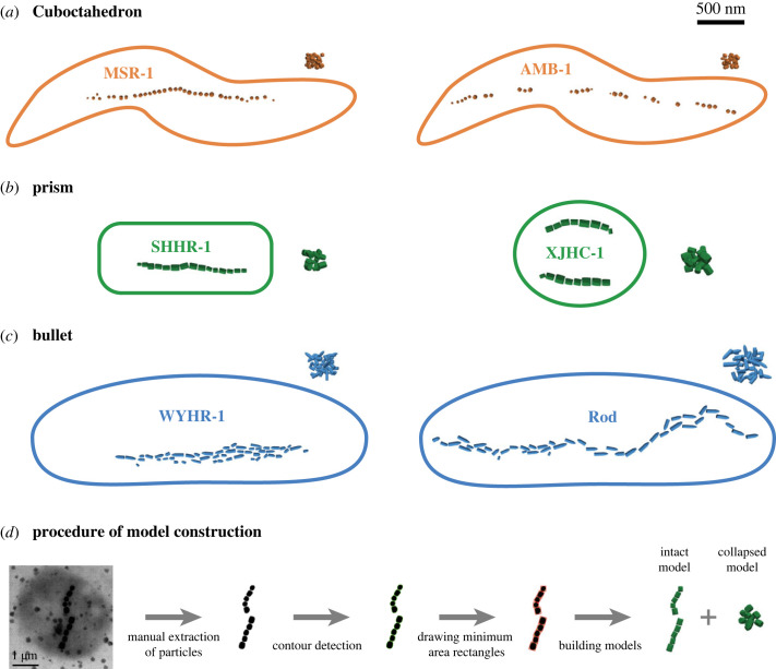
Micromagnetic model constructions of intact biogenic magnetite chains with three crystal forms and the corresponding chain collapse models (a–c), and an example of model construction procedures based on microscopic images (d). Published transmission electron microscopic (TEM) images of six typical magnetotactic bacteria were used to create these models: (a) MSR-1 [3] and AMB-1 [11] for cuboctahedron magnetosomes, (b) SHHR-1 [11] and XJHC-1 [30] for prism magnetosomes, (c) WYHR-1 [31] and a rod MTB [2] for bullet magnetosomes. Green curves and red rectangles in (d) represent detected particle contours and recognized minimum area rectangles using the OpenCV-Python library, respectively.

