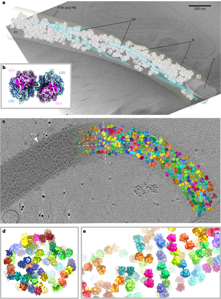
Fig. 1. Structure and organization of the S. lophii ribosome dimer in situ.
a, A segmented tomogram of a PT, showing ER (transparent blue), a vesicle (V, transparent magenta) and ribosomes (R, white). The PT wall (PTW) and plasma membrane (PM) are transparent grey. b, A subtomogram average of the S. lophii ribosome dimer (composite of two half-dimers) at 10.8 Å resolution, showing the SSU in shades of pink and the LSU in shades of blue. c, Organization of ribosome dimers inside a PT showing the original tomographic slice on the left and subtomogram averages of ribosome dimers placed back into the tomogram on the right (various colours). d,e, Cross-sections through the PT from areas indicated by the dotted lines (designated d and e) in c.

