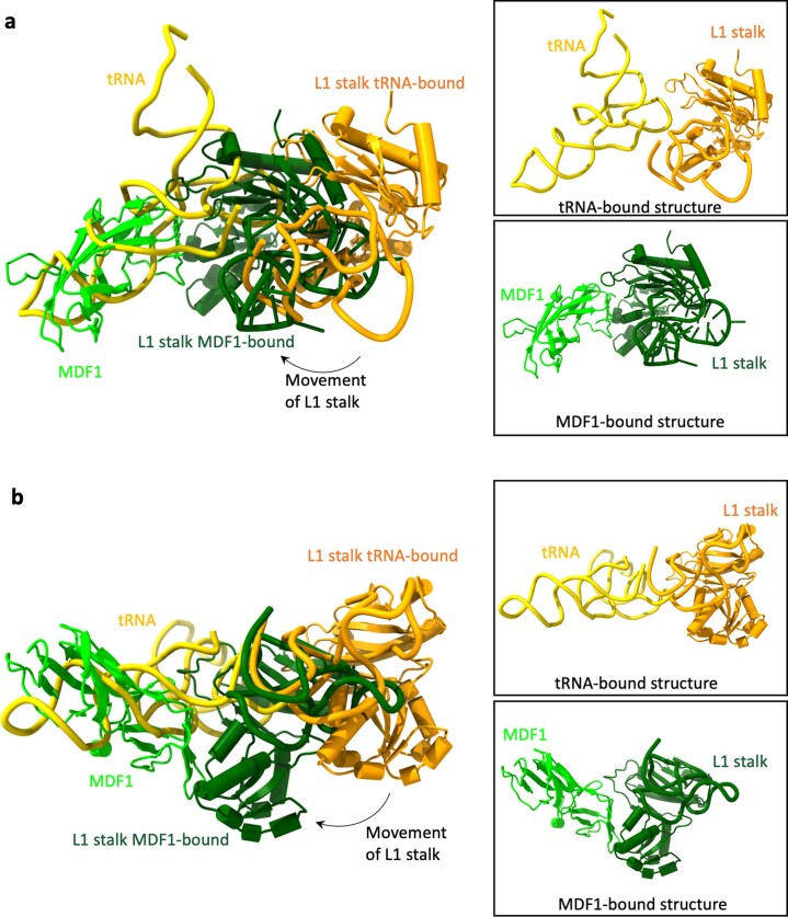
Extended Data Fig. 9. Comparison of the S. lophii L1 stalk position in the tRNA-bound and MDF1-bound states.
a, b Two different views of the S. lophii L1 stalk in an overlay of the tRNA-bound and MDF1-bound states. tRNA shown in yellow with corresponding L1 stalk (consisting of the uL1 protein and rRNA) in orange (from the monomeric S. lophii ribosome structure, 7QCA). MDF1 in lime with corresponding L1 stalk (consisting of the uL1 protein and rRNA) in dark green (from the dimeric S. lophii ribosome structure, 8P5D). Boxed figures show the aligned tRNA-bound and MDF1-bound states alone for each view to clarify the difference observed between the positions of the L1 stalk.

