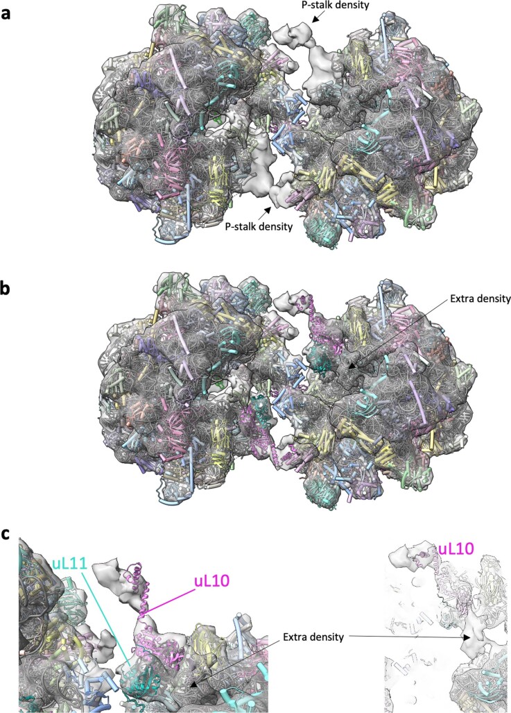
Extended Data Fig. 10. The P-stalk density of the 100SS. lophii ribosome.
a S. lophii dimer model (PDB 8P60) (cylinders and stubs) and 14.3 Å map (transparent grey; EMD-17457) showing additional density for the P-stalk (black arrows). b, c Alphafold2 models of the S. lophii proteins uL10 (pink ribbon) and uL11 (cyan ribbon) were modelled into the dimer map guided by the structure of the porcine ribosome (3J7P23) - one of the few ribosome structures with a partially ordered P-stalk. An area of extra density was observed in a region where elongation factors have been observed to bind, but the density was not clear enough to model any proteins.

