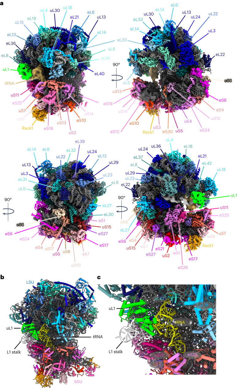
Fig. 2. Single particle structure of the S. lophii 70S ribosome at 2.3–2.9 Å resolution.
a, Various views of the ribosome, showing the protein chains of the LSU in shades of blue, the SSU in shades of red and the rRNA in grey. Subunit names are indicated. b, Atomic model of the ribosome with uL1 in lime green and tRNA in yellow. c, Magnified view of the E site of the ribosome showing deacetylated tRNA in yellow, L1 rRNA in white and protein uL1 in lime green. The tRNA interacts with protein uS7 of the SSU, proteins eL42 and uL1 of the LSU and rRNA.

