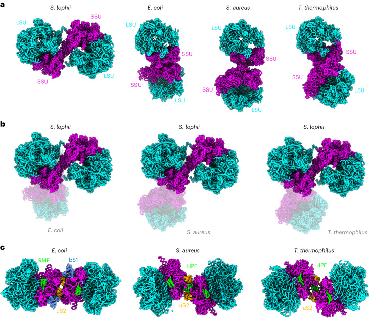
Fig. 4. Distinct dimer architecture in eukaryotes and bacteria.
a, Atomic models of S. lophii and various bacterial species shown side by side (licorice representation). The 70S ribosomes (half dimers) indicated by a white star (*) are in the same orientation. b, Hibernating ribosomes from three bacterial species (transparent) superimposed with that of S. lophii (opaque), highlighting distinct dimer architectures. c, Bacterial dimer interfaces with key subunits highlighted. Note that while in S. lophii and bacteria the dimer interface is established via the small ribosomal subunit, the exact location differs. In S. lophii, the dimer interface is established via the ribosomes’ beaks, while it is formed near uS2 in bacteria and mediated by the hibernation factors RMF/bS1 in E. coli and HPF in S. aureus and T. thermophilus.

