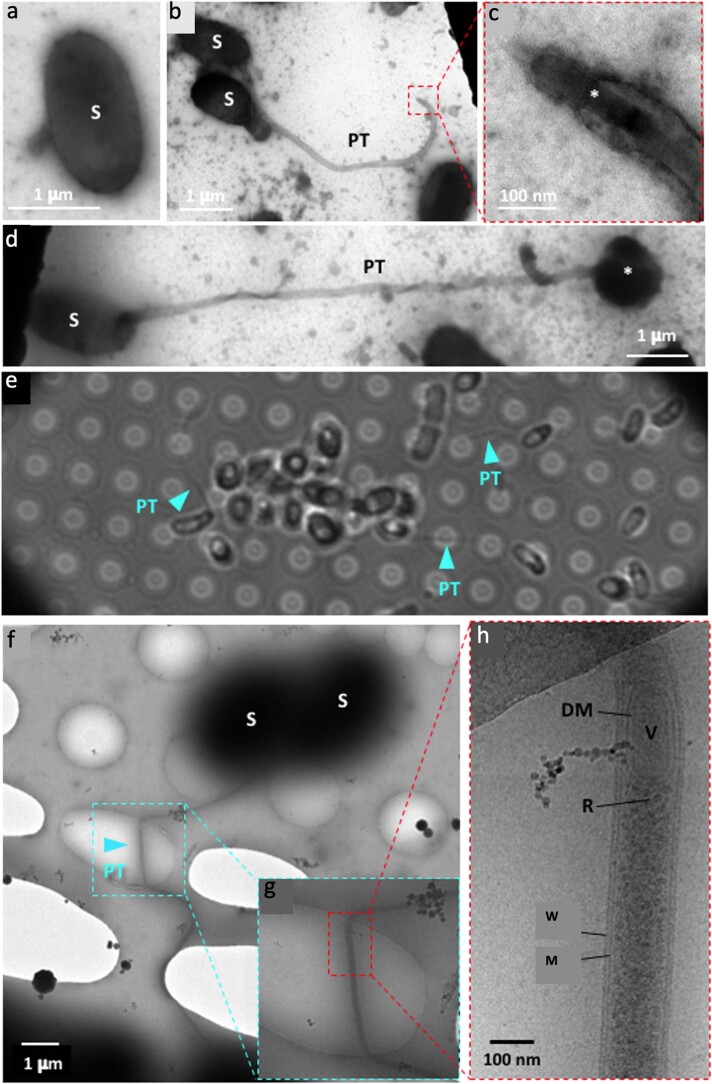
Extended Data Fig. 1. Electron microscopy of microsporidia germinated on EM grids.
a–d Negative stain EM of a a dormant spore (S), b a germinated spore with polar tube (PT) extended, c the tip of a PT with sporoplasm (*) passing through at high magnification and d a germinated spore post transfer, with sporoplasm egressing terminally (*). e Bright field light microscopy showing that microsporidia can be germinated on holy carbon (Quantifoil) grids. PTs are indicated with blue arrowheads. f-h CryoEM of microsporidia germinated on Multi-A Quantifoil grids, showing a PT emerging from a spore at three different magnifications. The PTs span Quantifoil holes, which is an important requirement for cryoET. g, h Higher magnification images of dashed area in f. h The PT is confined by a membrane (M) and proteinaceous PT wall (W). Transported content is visible. (R) ribosomes, (V) vesicle and (DM) double membrane. These images are representative of hundreds of micrographs collected from multiple (5) sample preparations.

