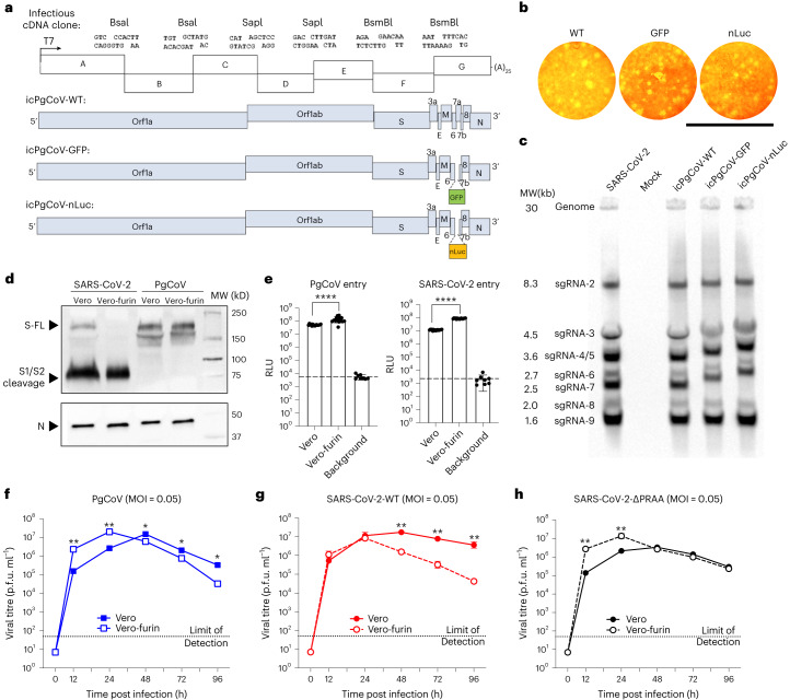
Fig. 1. Generation and characterization of recombinant PgCoVs.
a, Schematic design of PgCoV GD1 infectious cDNA clone. b, Plaque formation of the three recombinant PgCoVs. Scale bar, 8 mm. c, Northern blot analysis of genomic and subgenomic mRNAs isolated from SARS-CoV-2 and PgCoV infected cells at 24 h. d, Western blot analysis of semi-purified PgCoV and SARS-CoV-2 virions cultured in Vero or Vero-furin cell lines identified as full-length (FL), S1/S2 cleaved spike protein (S) and nucleocapsid protein (N). Samples were loaded on the basis of an equal amount of the N protein; this western blot was repeated twice with the same result. e, Efficient entry of PgCoV-nLuc and SARS-CoV-2-nLuc recombinant viruses into Vero-81 and Vero-furin cells at an MOI of 2. After 1 h infection, viruses were removed and cells were treated with neutralization antibodies to minimize secondary rounds of infection. The RLU representing the nLuc expression level was measured at 12 h post infection (n = 8 replicates per group, data are mean ± s.d.) and analysed using unpaired t-test; ****P < 0.0001. f–h, Multistep growth curves of PgCoV-WT (f), SARS-CoV-2-WT (g) and SARS-CoV-2-∆PRRA (h) in Vero and Vero-furin cells.

