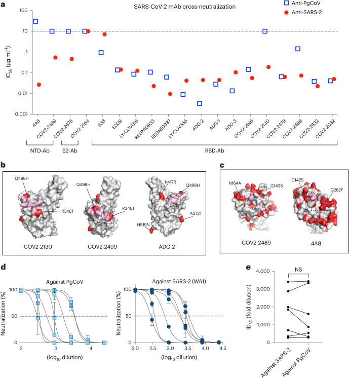
Fig. 5. SARS-CoV-2-specific antibodies neutralized PgCoV infection in vitro and in vivo.
Neutralization assays were performed using SARS-CoV-2 nLUC and PgCoV-nLUC recombinant viruses and panels of neutralizing monoclonal antibodies and convalescent patient sera. a, Summary of IC50 values from a panel of SARS-CoV-2 neutralizing antibodies. b, RBD antibody binding sites (pink) impacted by PgCoV natural variation (red). c, NTD antibody binding sites (pink) impacted by PgCoV natural variation (red). Binding sites in b and c were annotated on the basis of a SARS-CoV-2 spike structure (PBD ID: 6zp7). d, COVID-19 patient sera 8-point neutralization curves against SARS-CoV-2 (WA1) and PgCoV; each sample was run in triplicate in the assay, data are mean ± s.d. e, COVID-19 ID50 neutralizing titres against SARS-CoV-2 and PgCoV. Values were analysed using paired t-test; NS, P > 0.05.

