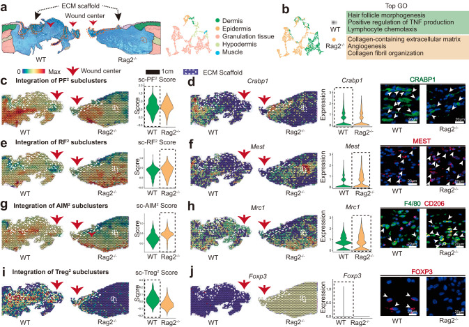
Fig. 6. Spatial atlas of cell microenvironment around biomaterials of immunodeficient mice.
a The anatomical structure of each sample. b Gene enrichment analysis between WT and Rag2-/- group. c Spatial feature plot and violin plot showing the distribution and expression level of integrated PF2 subcluster in tissue sections. d Spatial feature plot and violin plot showing the distribution and expression level of Crabp1 (marker gene of PF2) in tissue sections; Representative IF images of stained PF2 (CRABP1+), white arrowheads showing the CRABP1+ cells. e Spatial feature plot and violin plot showing the distribution and expression level of integrated RF2 subcluster in tissue sections. f Spatial feature plot and violin plot showing the distribution and expression level of Mest (marker gene of RF2) in tissue sections; Representative IF images of stained RF2 (MEST+), white arrowheads showing the MEST + cells. g Spatial feature plot and violin plot showing the distribution and expression level of integrated AIM2 subcluster in tissue sections. h Spatial feature plot and violin plot showing the distribution and expression level of Mrc1 (marker gene of AIM2) in tissue sections; Representative IF images of stained AIM2(F4/80 +CD206+), white arrowheads showing the F4/80 +CD206+ cells. i Spatial feature plot and violin plot showing the distribution and expression level of integrated Treg2 subcluster in tissue sections. j Spatial feature plot and violin plot showing the distribution and expression level of Foxp3 (marker gene of Treg2) in tissue sections; Representative IF images of stained Treg2 (FOXP3+), white arrowheads showing the FOXP3+ cells.

