Figure 2.
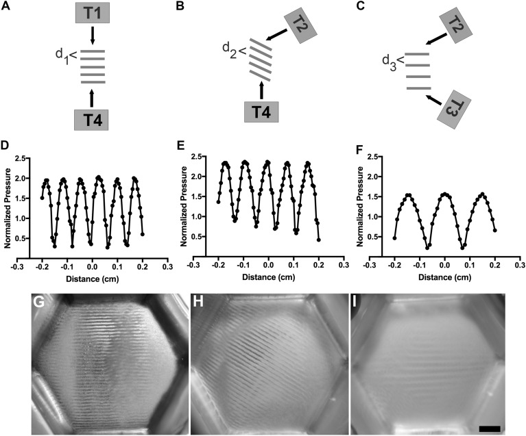
USWF-patterning of microparticles in vitro. (A–C) An USWF is generated at the intersection of two ultrasound fields. The angle of intersection of the fields influences the spacing and orientation of nodal planes (Eq. 3). Illustrated are beams intersecting at 180° (A), 120° (B), and 60° (C). ‘T’ denotes placement of transducers. (D–F) A membrane hydrophone was used to measure axial distributions of pressure in an USWF for angles of 180° (D), 120° (E), and 60° (F). Pressure measurements were normalized to the average of the free-field pressure measured from each active transducer. (G–I) Sephadex particles (10–40 µm diameter) were suspended in 1% (w/v) agarose solutions and exposed to an USWF generated by two transducers. Shown are images of microparticle patterning using 1-MHz ultrasound beams intersecting at 180° (G), 120° (H) and 60° (I). Images are representative of n = 3–4 gels fabricated over 4 independent experiments. Scale bar = 5 mm.

