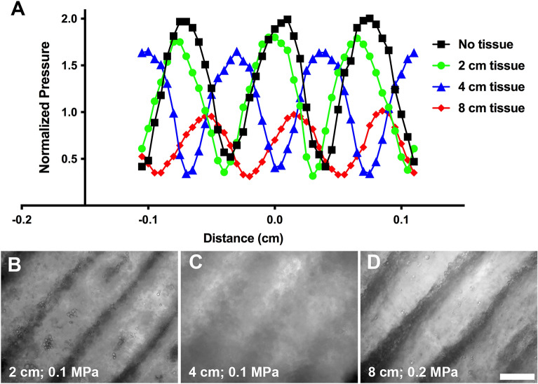
Figure 3.
USWF formation and acoustic patterning through intervening tissue. (A) Hydrophone measurements were used to determine axial spatial distributions of pressure in the free field in the presence of no tissue (black squares), or 2 cm (green circles), 4 cm (blue triangles), or 8 cm (red diamonds) of total intervening porcine muscle tissue. USWF pressure measurements were normalized to the average free field pressure measured from each active transducer. (B–D) Sephadex particles (10–40 µm diameter) suspended in collagen solutions (2 mg/mL) were exposed to 1-MHz USWFs at 0.1 (B,C) or 0.2 (D) MPa for 15 min. Collagen solutions polymerized during USWF exposure, thereby maintaining particle patterning after sound deactivation. Shown are representative microscopy images of Sephadex particles (phase dense) patterned using 1-MHz ultrasound beams intersecting at 180° in the presence of 2 (B), 4 (C), or 8 (D) cm of total intervening tissue. Images are representative of 3 gels per exposure condition, fabricated over 3 independent experiments. Scale bar = 500 µm.

