
Figure 1: Rapid HNF4α nuclear to cytoplasmic translocation after non-regenerating dose of APAP.
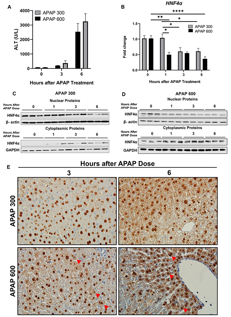
(A) Serum ALT levels (B) qPCR analysis of HNF4α mRNA at various time points after administration of 300 mg/kg and 600 mg/kg APAP in C57BL/6J mice. (C) Western blot analysis of HNF4α using nuclear and cytoplasmic extracts showing nuclear to cytoplasmic translocation of HNF4α after 300 mg/kg dose of APAP and (D) 600 mg/kg dose of APAP. (E) Representative photomicrographs of HNF4α immunohistochemistry staining at 3 and 6 h after 300 mg/kg and 600 mg/kg dose of APAP. Arrows pointing to cytoplasmic HNF4α staining. Original magnification, 600X; * Indicates significant difference at, *=P<0.05, **=P<0.01, ****=P<0.0001