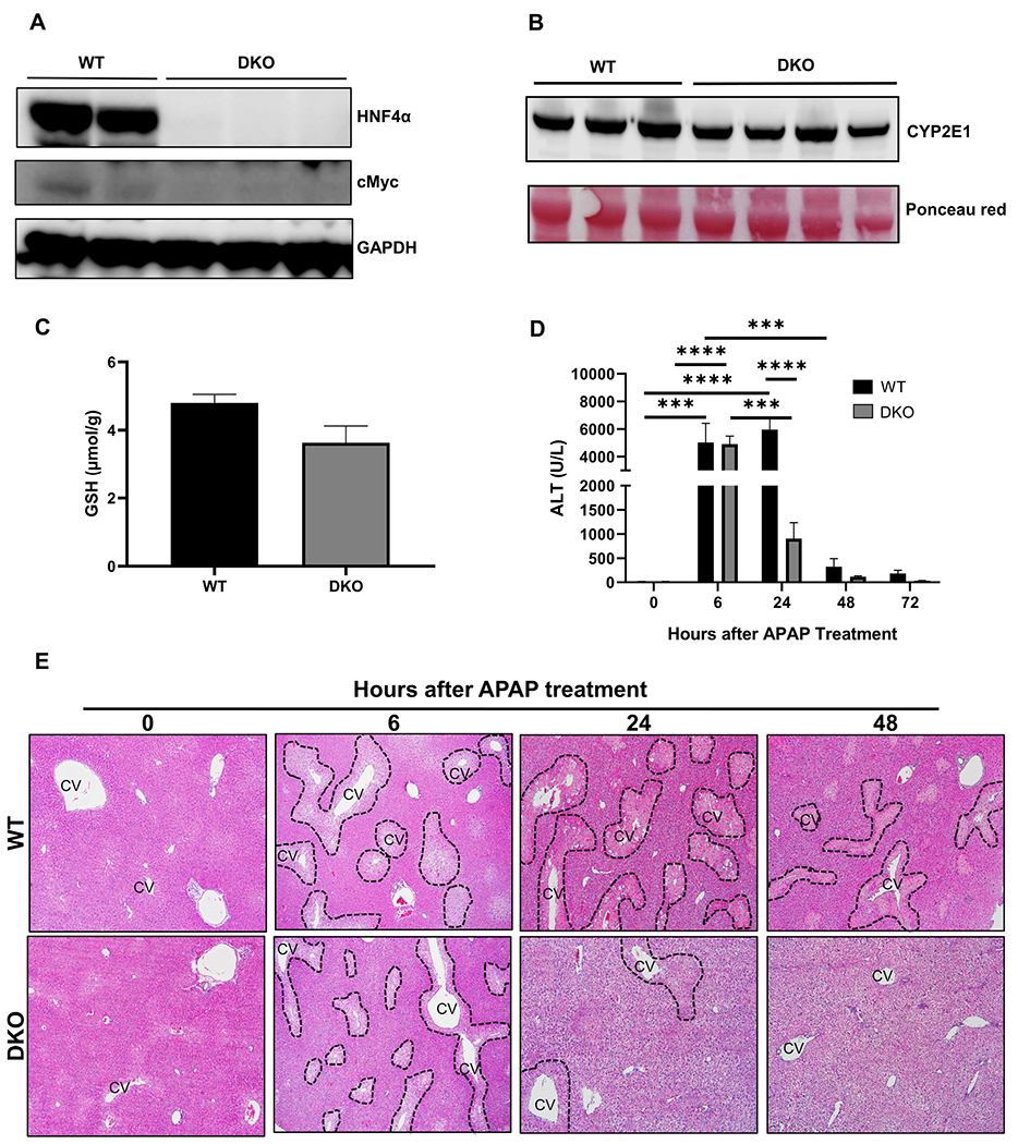
Figure 5: cMyc deletion in HNF4α-KO mice reduced APAP induced liver injury.

(A) Western blot analysis of HNF4α and cMyc showing deletion of HNF4α and cMyc in DKO mice (B) Western blot analysis of hepatic CYP2E1 and (C) Hepatic GSH levels in WT and DKO mice at 0 h time point. (D) Serum ALT levels of WT and DKO mice treated with APAP 300 mg/kg dose at various time points. (E) Representative photomicrographs of H&E-stained liver sections. Dotted lines demark area of necrosis. CV, central vein; Original magnification, 200X; * Indicates significant difference at, **=P<0.01, ***=P<0.001, ****=P<0.0001.
