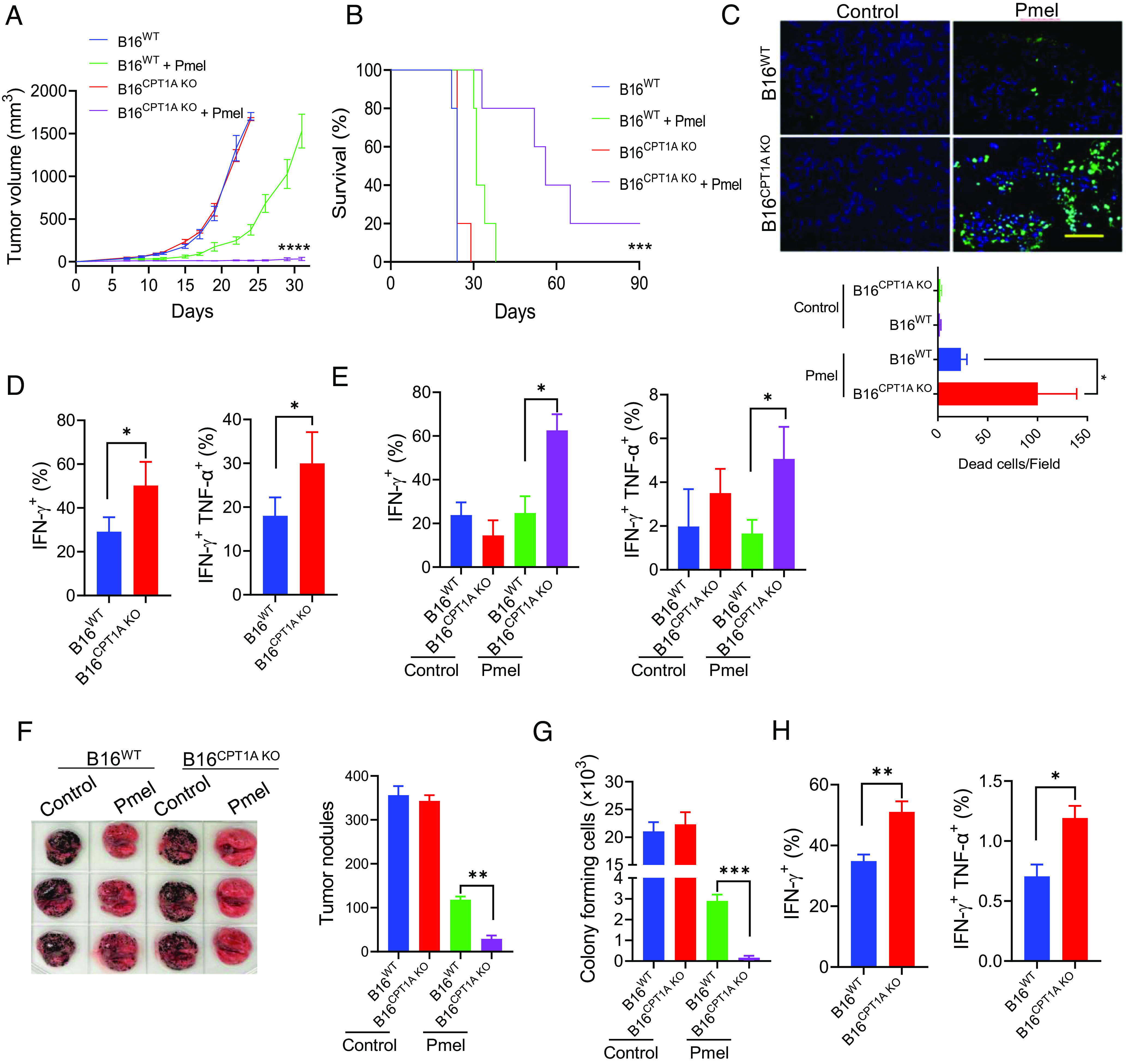
Fig. 3.

Lack of CPT1A renders metastatic melanoma susceptible to T cell therapy. C57BL/6 mice (n = 5) were injected with B16WT or B16CPT1A KO tumor cells (2 × 10⁵, s.c.) on day 0. Mice received two doses of Pmel T cells on days 4 and 7 (107 cells, i.v.) or left untreated. Tumor growth (A) and animal survival (B) were followed. (C) Tumor-bearing mice were treated with Pmel T cells on day 17 post-tumor inoculation; tumor tissues were collected on day 21 and subjected to TUNEL assays. The frequencies of IFN-γ+ or IFN-γ+ TNF-α+ Pmel cells (D) or IFN-γ+ or IFN-γ+ TNF-α+ endogenous CD8+ T cells (E) in tumors were assessed by intracellular cytokine staining and flow cytometry analysis (gating on total cells in tumor tissue). (F) C57BL/6 mice (n = 5) were established with experimental lung metastases by inoculating B16WT or B16CPT1A KO tumor cells (1 × 10⁵ cells, i.v.) on day 0. Mice received Pmel T cells (107 cells, i.v.) on day 8 or left untreated. Tumor nodules in the lungs were quantified on day 19. (G) Colony-formation assays were performed using lung cell suspensions to examine the frequency of metastatic cells. (H) The frequency of IFN-γ+ and IFN-γ+TNF-α+ CD8+ T cells in the lungs was analyzed by flow cytometry analysis (gating on total cells in lung tissues). Data are representative of three independent experiments. *P < 0.05. **P < 0.01. ***P < 0.001. ****P < 0.0001.
