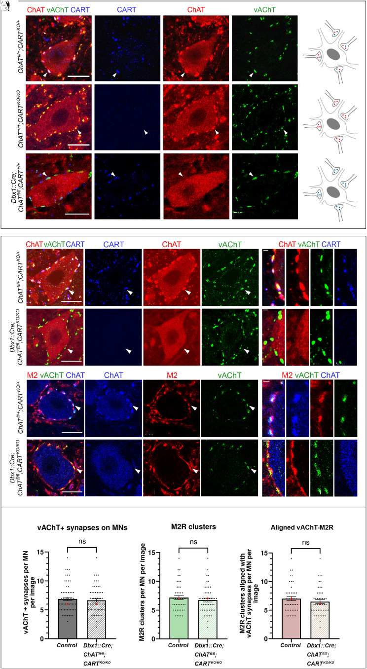
Fig. 5.
Gross anatomy of C boutons remains unchanged after genetic elimination of CART and ChAT from the synapse. (A–Aiii) C boutons on somata of motor neurons of wild-type mice are marked by CART (blue) and the cholinergic markers ChAT (red) and vAChT (green). (B) Schematic representation of a motor neuron receiving C boutons containing acetylcholine (red) and CART (blue). (C–Ciii) ChAT (red) and vAChT (green) expression are not disrupted in C boutons of CARTKO/KO mice. (D) Schematic representation of a motor neuron receiving C boutons containing acetylcholine (red) only. (E–Eiii) CART (blue) and vAChT (green) expression are not disrupted following the genetic deletion of ChAT in V0c interneurons. (F) Schematic representation of a motor neuron receiving C boutons containing CART (blue) only. (G–Giii) CART (blue), ChAT (red), and vAChT (green) are present in C boutons of herterozygous ChATfl/+;CARTKO/+ mice. The white arrowhead points to one representative triple-positive C bouton. (H–Hiii) High-magnification image showing the colocalization of CART (blue), ChAT (red), and vAChT (green) in individual C boutons. (I–Iiii) Despite the conditional knockout of ChAT and the complete knockout of Cart in Dbx1::Cre;ChATfl/fl;CARTKO/KO mice, the presynaptic components of C boutons remain intact, as evident by the presence of vAChT (green). The white arrowhead points to one CART−/ChAT− C bouton that expresses vAChT. (J–Jiii) High-magnification image showing the presence of vAChT (green) in individual C boutons that lack both ChAT and CART. (K–Kiii) The ChAT+/vAChT+ C boutons (blue and green respectively) on motor neurons of control ChATfl/+;CARTKO/+ mice are in close apposition to M2 muscarinic receptor clusters (red). The white arrowhead points to a ChAT+/M2+/vAChT+ C bouton. (L–Liii) High-magnification image showing the alignment of the postsynaptic M2 muscarinic receptor (red) with the ChAT+/vAChT+ presynaptic part of the synapse (red and green respectively). (M–Miii) Postsynaptic clustering of M2 receptors (red) with presynaptic vAChT (green) is unaltered in Dbx1::Cre;ChATfl/fl;CARTKO/KO double KO mice. The white arrowhead points to a vAChT+/M2+ synapse. (N–Niii) High-magnification image showing the alignment of M2 postsynaptic muscarinic receptors (red) with the vAChT+ (green) C boutons in Dbx1cre/+;ChATfl/fl;CARTKO/KO mice. (O–Q) No change was observed in the number of vAChT+ terminals (P = 0.6730, two-tailed), M2 muscarinic receptor clusters (P = 0.4402, two-tailed) or their alignment (P = 0.2880, two-tailed) despite the simultaneous elimination of ChAT and CART in C boutons. The bar charts represent the mean number of synapses per motor neuron with superimposed data points representing the data distribution. Data are presented as mean values (±SEM) and were combined from different sexes as no sex-dependent differences were observed. The median is also presented as a red dot superimposed in each graph. For panels O–Q “image” corresponds to optical thickness of 1,208 μm. [Scale bars, 20 μm, (A–M), 1 μm (H–N).]

