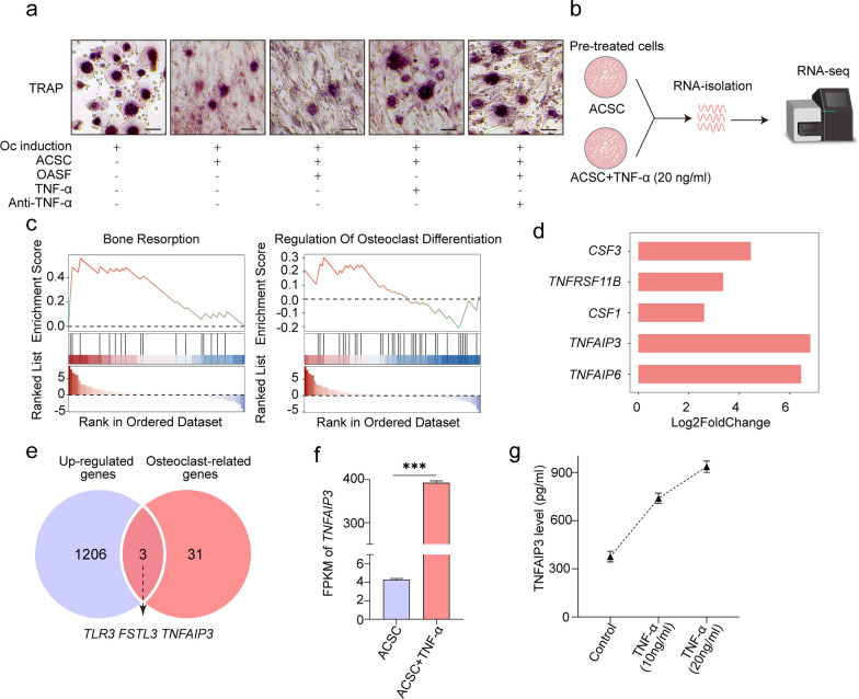
Fig. 4.
The function and expression of TNFAIP3 in ACSCs. a Representative TRAP staining images of osteoclasts during in vitro osteoclast formation in the presence of 5% OASF, 20 ng/mL TNF-α or 250 ng/mL anti-human TNF-α neutralizing antibody showing that TNF-α and OASF (containing TNF-α) suppress osteoclast formation. Scale bar, 50 μm. b Schematic overview of the RNA sequencing workflow. ACSCs were treated with 20 ng/mL TNF-α for 3 d, followed by RNA isolation, cDNA library construction and high-throughput sequencing (n = 3). c GSEA enrichment plots showed that ACSCs were more actively involved in osteoclast-related biological processes after TNF-α stimulation. d Bar plot showing the differential expression of osteoclast-related genes upon TNF-α stimulation. e Venn diagram indicated upregulated osteoclast-related DEGs in TNF-α stimulated ACSCs. f Gene expression of TNFAIP3 in ACSCs treated without or with 20 ng/mL TNF-α. g ELISA quantification of TNFAIP3 in the culture supernatant of ACSCs treated without or with TNF-α (10 ng/mL or 20 ng/mL) for 3 days (n = 3)

