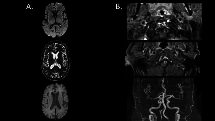
Fig. 2.
Brain MRI of one patient from the OSA group with evidence of intracranial vessel gadolinium enhancement without vascular stenosis nor any parenchymal lesion. Legend: A from the top to the bottom, we show brain axial slides of diffusion-weighted imaging (DWI, top), T2-weighted (middle) and susceptibility-weighted images (SWI, bottom) brain MRI sequences. They do not find any brain parenchymal damage. B from the top to the bottom, we show axial slide of dynamic 3D contrast-enhanced MR angiography (MRA) of the neck vessels at the vertebral arteries levels (top) and at the basilar artery level (middle) as well as 3D time-of-flight (TOF) MRA of the intracranial vessels (bottom). The first two images show gadolinium contrast enhancement of both vertebral arteries and basilar artery, without any vascular stenosis (TOF, third image)

