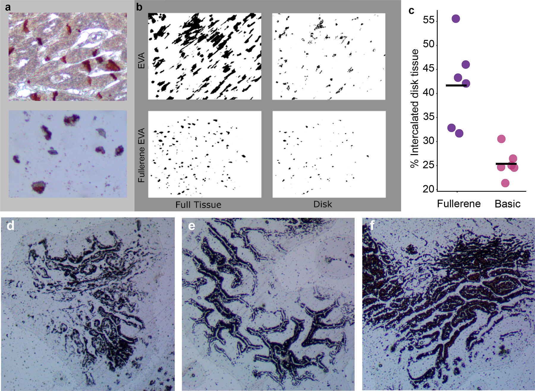
Figure 3.

A comparison between fullerene-impregnated EVA membranes and unimpregnated membranes. (A) Human heart slides, stained for intercalated discs (N-cadherin) and cut at 4 μm. were used. Top panel demonstrates the staining pattern for intercalated discs and bottom panel an EVA membrane. (B) Binary color area images of a postdissected unimpregnated EVA membrane showing the full tissue captured (top left) vs the disk region within the collected tissue (top right). Similar images of a postdissected fullerene-impregnated EVA membrane (bottom left) demonstrating improved capture specificity with less full tissue relative to captured disk material (bottom right). (C) An ImageJ pixel count across multiple images (n = 12) demonstrated increased specificity in capturing intercalated disc tissue with fullerene-impregnated EVA membranes. (D-F) 6%. 8%. and 10% EVA membranes showing capture of AE1/AE3’ epithelial cells, with the cleanest pattern seen with 8% EVA. EVA. ethylene vinyl acetate
