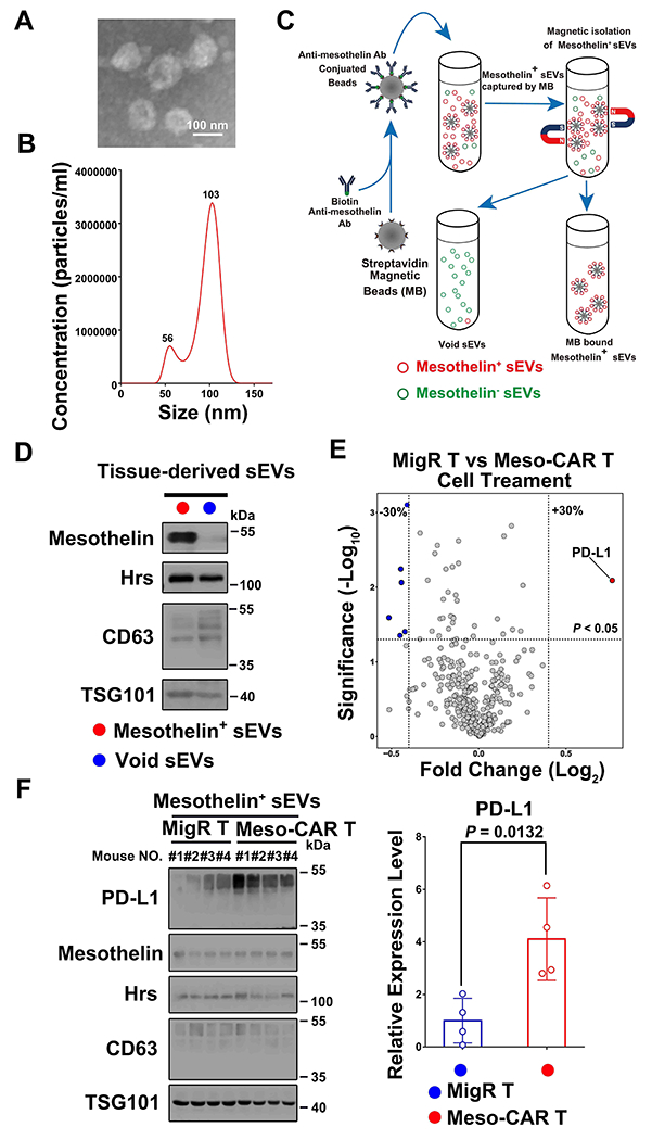
Figure 1. PDAC cells release sEVs that carry high levels of PD-L1 in response to Meso-CAR T treatment.

A, Representative electron microscopic image of sEVs isolated from tumor tissues. Scale bar, 100 nm. B, Characterization of sEVs purified from PDAC tumor tissues by nanoparticle tracking. The X-axis represents the diameters of the isolated vesicles; the Y-axis represents the concentration of isolated vesicles (particles/ml). C, Isolation of Mesothelin+ sEVs from PDAC tumor tissue-derived sEVs by magnetic beads. See Materials and Methods for details. D, Characterization of Mesothelin+ sEVs and remaining (“Void sEVs”) sEVs by western blotting. The expression levels of mesothelin and exosome marker proteins (Hrs, CD63 and TSG101) in indicated sEVs are shown. An equal amount of sEV proteins from the different fractions was loaded on the gel. E, Volcano plot of RPPA data displaying the pattern of protein expression comparing sEVs derived from PDAC tumors treated with MigR T cells or Meso-CAR T cells. The dotted horizontal line represents a significance level of P < 0.05, while dotted vertical lines represent differential expression differences of ± 30% (n=3). Each point represents the difference in the expression of each protein in the indicated sEVs. PD-L1 level was significantly higher in the sEVs from tumors with Meso-CAR T treatment than those with MigR T cell treatment. F, Western blot analysis of PD-L1, mesothelin and exosome marker proteins (Hrs, CD63 and TSG101) in purified tumor cell-derived Mesothelin+ sEVs from PDAC tumors with MigR T cell or Meso-CAR T cell treatment. All lanes were loaded with equal amounts of proteins. Quantification of the levels of PD-L1 in the sEVs is shown to the right (n=4). Data represent mean ± s.d. (n=3 or as indicated). Statistical analysis was performed using two-sided unpaired multiple t-test (E) or two-sided unpaired t-test (F).
