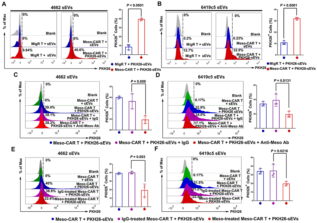
Figure 3. Mesothelin mediates the interaction between TEVs and CAR T cells.

A and B, Representative images of flow cytometry of MigR and Meso-CAR T cells after incubation with PKH26-labeled 4662 cell-derived sEVs (A) and 6419c5 cell-derived sEVs (B). The percentages of sEV-bound MigR and Meso-CAR T cells are shown at the right. C and D, Representative images of flow cytometry of Meso-CAR T cells bound to PKH26-labeled-4662 cell-derived sEVs (C) and 6419c5 cell-derived sEVs (D) that were pre-treated with or without IgG or anti-mesothelin antibodies. The percentages of PKH26 positive Meso-CAR T cells are shown at the right. E and F, Representative images of flow cytometry of Meso-CAR T cells that bound to PKH26-labeled sEVs. Meso-CAR T cells were pre-treated with recombinant mesothelin and then incubated with PKH26-labeled sEVs derived from 4662 cells (E) and 6419c5 cells (F). The percentages of PKH26-positive Meso-CAR T cells are shown at the right. Data represent mean ± s.d. (n=3). Statistical analysis is performed using two-sided unpaired t-test (A, B) or one-way ANOVA analysis with Dunnett’s multiple comparison tests (C-F).
